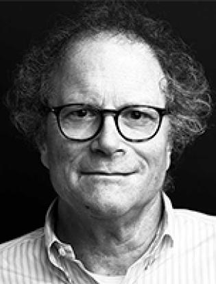
Michael Barish, Ph.D.
Duarte Cancer Center
Duarte, CA 91010
1980 - 1985, University of California, Los Angeles, Los Angeles, CA Post-doc, Physiology
1980, Stanford University, Stanford, CA, Ph.D., Biological Sciences
1972, M.I.T., Cambridge, MA, S.B., Biology
2015 - present, Chair and Professor, Department of Stem Cell Biology and Regenerative Medicine, Beckman Research Institute of City of Hope, Duarte, CA
2001 - 2015, Chair, Department of Neurosciences, Beckman Research Institute of City of Hope, Duarte, CA
2000 - present, Professor of Neurosciences, Beckman Research Institute of City of Hope, Duarte, CA
1989 - 2004, Adjunct Professor (Asst., Assoc., Full), University of California, Irvine, CA
1995 - 2001, Associate Chair, Division of Neurosciences, Beckman Research Institute of City of Hope, Duarte, CA
1989 - 2000, Assistant and Associate Research Scientist, Division of Neurosciences, Beckman Research Institute of City of Hope, Duarte, CA
1995 - 1996, Associate Adjunct Professor, Department of Biology, Harvey Mudd College, Claremont, CA
1985 - 1989, Assistant Adjunct Professor and Assistant Professor in Residence, Department of Physiology and Biophysics, College of Medicine, University of California, Irvine, Irvine, CA
1980 - 1985, Postdoctoral Fellow, Department of Physiology, University of California, Los Angeles, Los Angeles, CA
1974 - 1980, Graduate Student, Department of Biological Sciences, Stanford University, Stanford, CA
1972 - 1974, Science Teacher, Germantown Friends School, Philadelphia, PA
Physiological studies of neural progenitor cell differentiation
At all stages of brain development, cells of the central nervous system express highly variable combinations of ion channels and neurotransmitter receptors. The immature neurons and neural progenitor cells (NPCs) that populate the brain early in development have physiological properties that are distinct from those of mature cells and that drive the activity-dependence of nervous system development. Fundamental aspects of brain development are regulated by activity, and the natural progression of maturational events is interrupted when neural activity is silenced.
In the developing hippocampus, almost all neurons are recruited into episodes of spontaneous synchronous electrical activity that would look like seizure in a different non-developmental context. Non-excitable cells such as glial cells and, most significantly, neuro progenitor cells (NPCs), respond to this activity because of proximity and specific interactions. We are studying the responses of these NPCs in postnatal mouse hippocampus (identified in living slices by nestin promoter-driven EGFP expression), using imaging and electrophysiological techniques, and immunofluorescence and confocal microscopy.
Focusing on nestin: EGFP+ NPCs in the dentate gyrus during episodes of activity, we observe that their Ca2+ responses are heterogeneous, varying with cell morphology and position as these cells migrate from the ventricular zone to the hilus and then to the granule cell layer (GCL) and subgranular zone (SGZ). This heterogeneity implies that there are systematic variations in ion channel, neurotransmitter receptor and/or innervation with position and morphology, and we are exploring this aspect of the biology of these cells, comparing these aspects of NPC phenotype to stage-specific markers of neural differentiation.
In a parallel study, we are examining the activity-dependence of Cl- transporter expression in NPCs and maturing neurons, because a common characteristic of immature neurons is the depolarizing nature of GABAA receptor activation, rather than hyperpolarizing as in mature neurons. This is due to elevated early expression of the inwardly-directed Na-K-2Cl cotransporter NKCC1. Subsequently, loss of NKCC1 and increased expression of the K-Cl cotransporter KCC2 reduces intracellular [Cl-] as neurons mature and GABAA-mediated inhibition becomes effective. We observe that expression of KCC2 in organotypic culture of hippocampal slice is sensitive to activity, and we are presently determining the signaling pathways linking activity and KCC2 expression, and investigating the consequences of altered KCC2 expression on structural rearrangements of the dentate gyrus, and on SGZ and GCL neuron differentiation.
Studies on the activity-dependence of tumor initiation and progression
Glioma and other brain cancers often present clinically as seizure, and studies using rodent models have shown that epileptic foci are found near but not in the tumor mass. Tumor cells, even if non-excitable, may be responsive elements in these activated networks, and electrically active neighboring host cells may be interacting with the tumors. This arrangement is reminiscent of that seen during brain development, in which non-excitable neuro progenitor cells are surrounded by, and react to, the activity of more mature neighbors. In our present efforts, done in collaboration with Drs. Karen Aboody (Hematology/HCT) and Carlotta Glackin (Molecular Medicine), we are approaching questions of initiation, progression and recruitment in precisely the same ways we are examining the activity-dependence of hippocampal development. Working with mice bearing intracranial transplants of human glioma lines, we have observed pleiotropic responses in the host brain to the presence of tumor cells, including a notable migration of endogenous NPCs towards and into the tumor mass.
Because epileptic foci may be in the normal brain surrounding the tumor mass, and spontaneous electrical activity during early development resembles seizure, in one series of experiments we are asking if the presence of glioma cells induces developmental regression in the physiological properties of surrounding neurons. U251 glioma cells are implanted to the cortex of mature mouse brains and allowed to expand for three weeks. We observe that human U251 tumor cells express NKCC1 and not KCC2, consistent with a relatively undifferentiated tumor phenotype. Except near the leading invading edge of the tumor, host mouse neurons show KCC2 immunoreactivity, consistent with their mature status. Mouse cells adjacent to invading tumor cells exhibit reduced KCC2 expression, a change expected (in the absence of other compensatory changes) to increase excitability by decreasing the efficacy of GABAergic inhibition. While preliminary, these observations are consistent with our hypothesis, and in future experiments we will work with more developed tumors and examine expression of additional developmentally-regulated differentiation markers along with transporters, ion channels and neurotransmitter receptors.
Studies on signaling between HB1.F3 immortalized human neural progenitor cells and their glioma targets
HB1.F3 cells, an immortalized human NPC line, will be home to experimental glioma (U251 cells transplanted into mouse brain) when injected into the brain parenchyma or blood circulation (tail vein). There is great interest in using these neuro progenitor cells to deliver therapeutic agents or as cytotoxic cells themselves. Finding tumor cells in the brain is a complex task, involving extravasation, migration and tumor cell recognition. The signaling mechanisms responsible for each of these aspects of tumor homing are not well understood. In collaboration with the Aboody and Glackin laboratories, we have been examining the last stage of this process: HB1.F3-tumor cell signaling.
Recent results from the Aboody/Glackin group have implicated hepatocyte growth factor (scatter factor; HGF) as a soluble signal able to serve as a chemoattractant in a modified Boyden chamber assay. This effect was abrogated by inhibiting PI-3 kinase activity through siRNA knock-down of its p85α subunit. We tested for the presence of additional signaling processes mediating cell contact or short-range diffusion interactions by tracking HB1.F3 cell movement on a monolayer of U251 tumor cells. We compared the cumulative displacements of wild-type and p85α-KD HB1.F3 cells on tumor cells with those observed when they were on the fibronectin-coated glass surround. We observed that both wild-type and p85α-KD HB1.F3 cells showed significantly reduced displacement on the surround as compared to on-tumor cells, and that there was no significant difference between displacement of wild-type and p85α-KD HB1.F3 cells when they were on the surround (and therefore presumably exposed to the same soluble factors as well as to a common substrate). However, there was a significant difference in displacement of wild-type and p85α-KD HB1.F3 cells when they were on the tumor cells, which suggests that HB1.F3 cell contact with U251 cells activates an additional set of intracellular signaling mechanisms. Future experiments will utilize Ca2+ imaging and pharmacological and genetic manipulations of HB1.F3 and U251 cells to dissect the elements of this signaling. Identifying these signals will enable refinement and expansion of the therapeutic potential of HB1.F3 cells. It will be particularly interesting to examine the extent to which HB1.F3-U251 cell signaling recapitulates or diverges from the signals passing between the neuro progenitor cells of the rostral migratory stream in normal adult brain (“transient amplifying cells”) and the GFAP-expressing astrocytes that guide them.
1995 - 2002, Editorial Board, Journal of Physiology (London)
1994 - 1999, Visiting Researcher, Overseas Research Invitation Program, Department of Molecular & Cellular Neurosciences Section, Electrotechnical Laboratory, Tsukuba, Ibaraki, Japan
1989 - 1993, Established Investigatorship, American Heart Association (National Center)
1986, Faculty Research Award, UC Irvine, California College of Medicine Annual Fund
1980, Arthur C. Giese Award in Marine Biology, Hopkins Marine Station, Stanford University, CA
