Electron Microscopy and Atomic Force Microscopy Equipment
Overview
- AKTA Oligopilot Plus
- AKTA Pure Purifier
- PELCO Biowave Pro 36500 Laboratory Microwave System
- Bruker Multimode 8 Atomic Force Microscope
- Cressington 308 Coating System
- PELCO easiGlow 91000 Glow Discharge Cleaning System
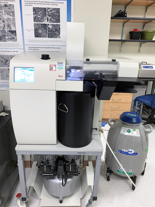
- Leica Electron Microscope Automatic Freeze Substitution System
- Leica Electron Microscope PACT2 High-Pressure Freezer
- FEI Quanta 200 Scanning Electron Microscope
- FEI Tecnai Twin 120kV Transmission Electron Microscope (TEM)
- FEI Vitrobot Mark IV
- FEI Vitrobot Mark IV
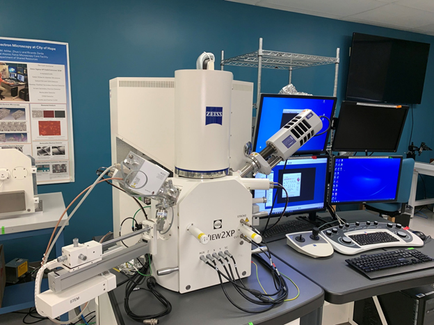
- Gatan 3View2XP In-chamber Microtome
- Gatan Sample Holder and Pumping Station
- GE AKTA Flux 6
- Leica Ultracut UCT Microtome for Ultrathin Sectioning
- Leica VT1000S Vibrating-blade Microtome
- Oxford X-MAX SDD X-ray Detector
- Polaron Critical Point Dryer
- PT-PC PC Controlled Ultramicrotome
- Spreading Coating Systems 6800 Spin Coater
- Zeiss Sigma VP Field Emission Scanning Electron Microscope (FE-SEM)
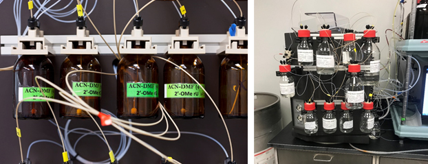
AKTA Oligopilot Plus
The Oligopilot plus synthesizer is utilized to synthesize quantities of oligonucleotides for development and early stage toxicology studies.
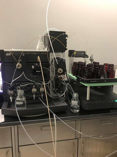
AKTA Pure Purifier
AKTApure is an antomated liquid chromatography system built for process development and small-scale biopharmaceutical manufacturing.
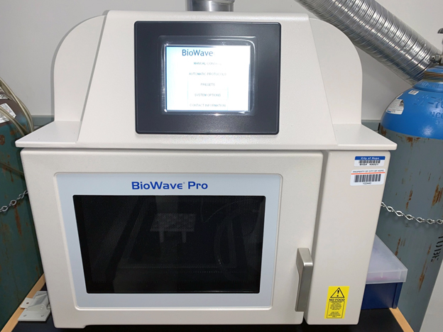
PELCO Biowave Pro 36500 Laboratory Microwave System
This instrument is available for specimen preparation allowing otherwise time-consuming sample preparation to be semi-automated and accomplished more quickly.
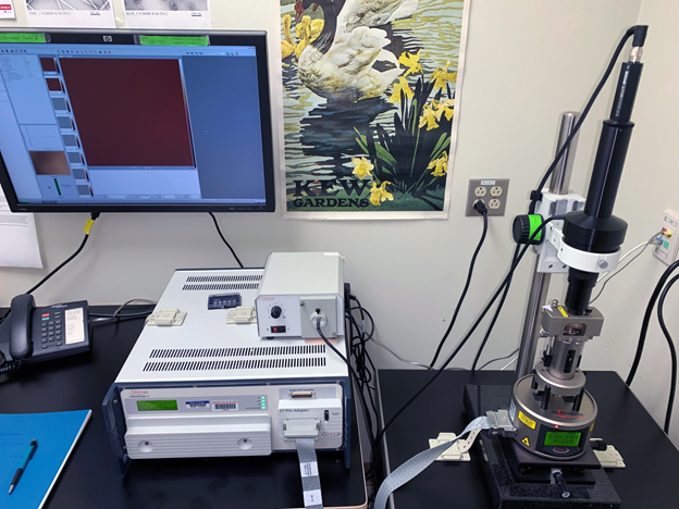
Bruker Multimode 8 Atomic Force Microscope
Bruker Multimode 8 AFM provides a means for imaging the surfaces of specimens using a physical probe and nearly all the major scanning-probe-microscopy imaging methods, including contact and non-contact atomic force, lateral force, tapping (air), magnetic force, electric force microscopy, and ScanAsyst (Peak Force Tapping) techniques. A cantilever holder allows for fluid scanning using tapping and force modulation modes. In addition, a ScanAsyst-HR high-speed scanner upgrade offers 20-times faster scanning with no loss of resolution.
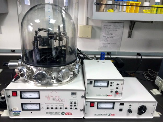
Cressington 308 Coating System
This coating system provides thermal evaporation of various metals. It is most often used for sputter coating SEM samples and depositing carbon onto TEM grids.
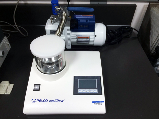
PELCO easiGlow 91000 Glow Discharge Cleaning System
For rendering TEM carbon support films hydrophilic (negatively charged).
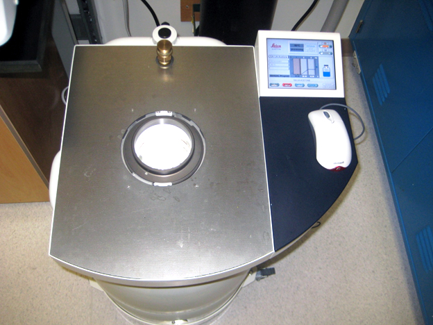
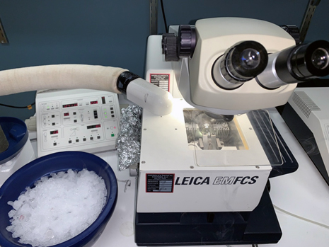
Leica Electron Microscope Automatic Freeze Substitution System
Freezing substitution is the follow-on procedure for high-pressure freezing. Specimens are fixed and resin-infiltrated at low temperatures providing higher quality structural preservation, including specimens suitable for immunolabeling for electron microscopy.
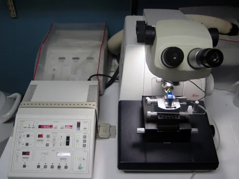
Leica Electron Microscope PACT2 High-Pressure Freezer
The HPF equipped with RTS allows ultra-rapid freezing of specimens. High-pressure freezing is superior to chemical fixation to preserve the native structure of cells and molecules. Antigenic sites in specimens are better preserved with HPF, opening greater possibilities for localization of molecules of interest within cells. Also available is a Leica Microbiopsy Transfer System (not shown), which provides the means for rapidly collecting microbiopsy specimens.
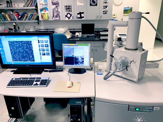
FEI Quanta 200 Scanning Electron Microscope
This SEM is a versatile, high-performance (tungsten filament) scanning electron microscope with three operating modes (high-vacuum, low-vacuum and ESEM) equipped with a motorized stage and detectors for secondary and backscattered electrons.
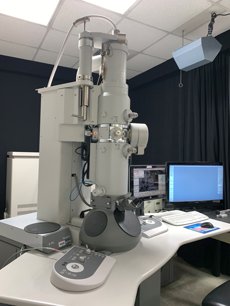
FEI Tecnai Twin 120kV transmission electron microscope (TEM)
This TEM provides several modes for directly imaging cell and macromolecular ultrastructure. The TEM can be used to examine negatively-stained specimens, cryo-specimens and sections of resin-embedded specimens. It is equipped with a Gatan Ultrascan 1000 2K CCD camera. A 70° tilt cryo-transfer holder enables frozen specimens to be imaged at temperatures below -170 °C. Software and hardware are available for acquiring and aligning tomography data for subsequent reconstruction and for single particle analysis.
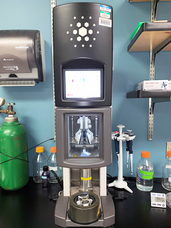
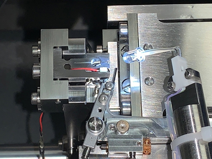
FEI Vitrobot Mark IV
This fully automated vitrification device for plunge-freezing of aqueous (colloidal) suspensions provides a powerful means for determining the 3D structure of macromolecules and macromolecular complexes in their natural state. Single-particle analysis and EM reconstruction of specimens prepared with the Vitrobot and analyzed by Cryo-EM can partner nicely with X-ray crystallography and NMR spectrometry in determining of molecular structures.
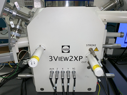
Gatan 3View2XP In-chamber microtome
The 3View2XP in-chamber microtome, when combined with the Zeiss Sigma VP FE-SEM, produces three-dimensional images in a fully-automated, high-throughput, high-resolution fashion. Data collected provide SBEM on the ultrastructure of cells and organelles and the opportunity for robust quantitative analyses at ultrastructural resolution. New updates include a focal charge compensation (Focal CC) device that dramatically improves image quality by eliminating charging effects.
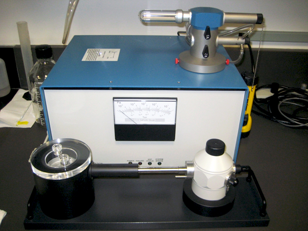
Gatan sample holder and pumping station
These are used for cryo-specimens in the TEM.
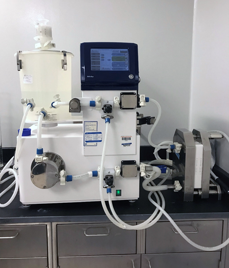
GE AKTA Flux 6
Semi-automated tangential flow filtration/cross flow filtration system for the concentration and diafiltration of purified oligonucleotides.
Leica Ultracut UCT Microtome for ultrathin sectioning
This versatile ultramicrotome is the workhorse instrument in the EM Core. It provides sections of resin-embedded specimens prepared by conventional and cryo methods. An FCS attachment allows low temperature ultrathin sectioning of biological samples at temperatures as low as -185°C following the method of Tokuyasu.
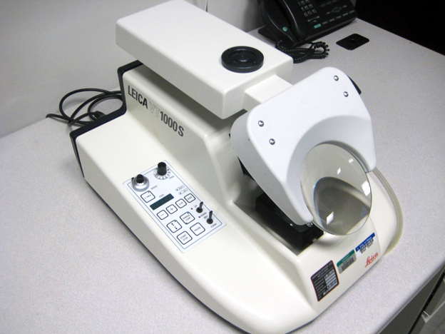
Leica VT1000S Vibrating-blade microtome, commonly called a vibratome
The vibrating razor blade in the vibratome provides relatively thick sections (~100 microns) suitable for specialized techniques of electron microscopy including immuno-EM.
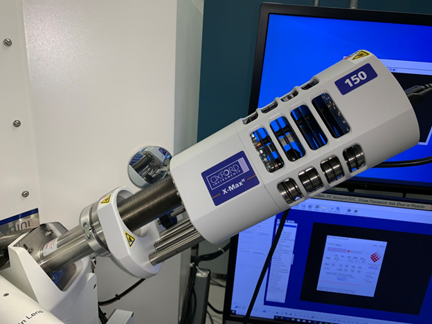
Oxford X-MAX SDD X-ray detector with a 150 mm2 window
This instrument is available for identifying and mapping elements within specimens.
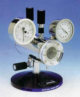
Polaron Critical Point Dryer
Critical point drying provides better preservation of specimens for scanning electron microscopy.
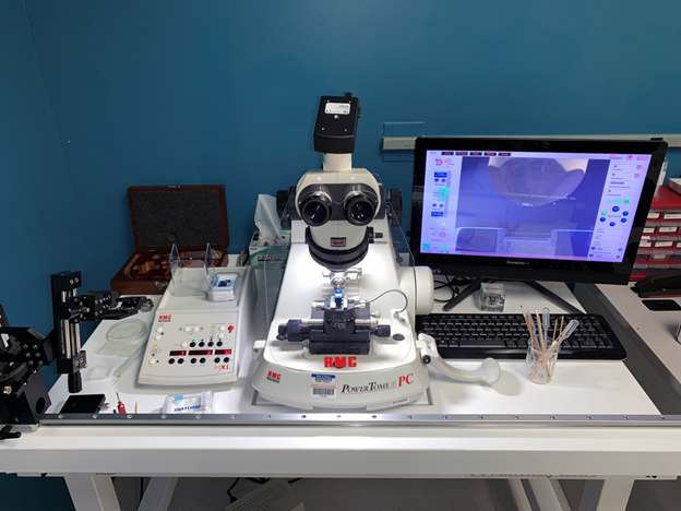
PT-PC PC Controlled Ultramicrotome from RMC Products, Boeckeler Instruments, Inc.
This new ultramicrotome is ATUMtome equipped and can be used for producing serial sections for viewing in the Sigma VP FE-SEM and producing 3D image data. The PT-PC also provides sections suitable for light and electron correlative microscopy.
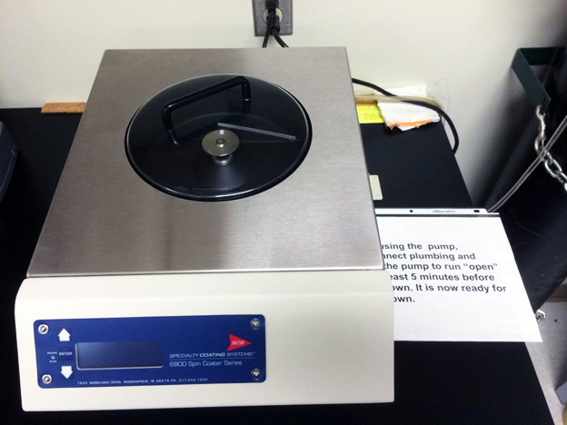
Spreading Coating Systems 6800 Spin Coater
For preparation of specimens for the scanning probe microscope.
Zeiss Sigma VP Field Emission Scanning Electron Microscope (FE-SEM) with Gemini column
This FE-SEM is equipped several detectors for secondary (SE) and backscattered (BSE) electrons. These include in-lens SE, variable pressure SE, Everhart-Thornley SE, BSE, and scanning transmission electron microscopy (STEM) detectors.