Light Microscopy/Digital Imaging Equipment
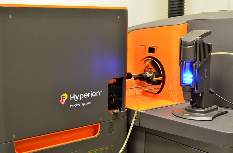
Hyperion Helios CyTOF
The Hyperion Imaging Mass Cytometer (IMC) is part of Fluidigm's line of CyTOF systems which allows for tissue samples to be “imaged” and for the masses of metals to be quantified in relation to the tissue morphology at a resolution of 1 square micron. The advantage of the system is that an investigator can label the tissue sample with as many as 50 different metal-tagged antibodies.
For more information please contact Light Microscopy Core personnel.
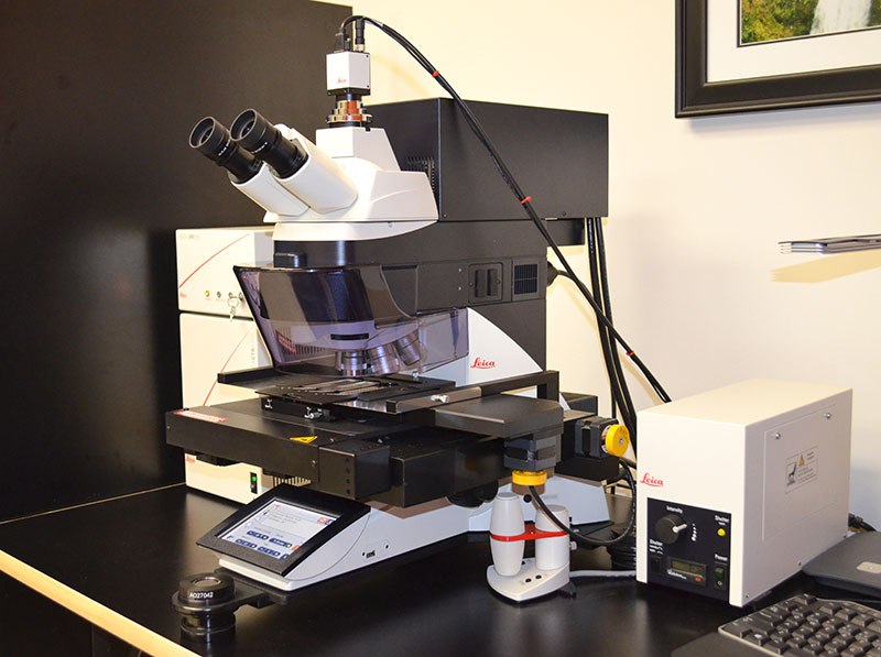
Leica LMD 7000
Widefield Visual Modes
- Bright field
- RGB Fluorescence: Metal Halide 120V
LASER
- 349nm (multiple adjustment parameters; solid state LASER)
Camera
- CC7000
Specimen Holders
- 3 Slides
- Petri Dish
- 18well slide
Collection Devices
- 0.2mL tubes
- 0.5mL tubes
- Universal Collection holder (with inserts)
Features
- Two Wacom Pen Screens; can draw shapes for cutting directly on monitor
- Can cut and collect multiple shapes into multiple collection devices automatically
- Live-Cell-Cutting (LCC) macro
- Collection tube inspection macro
- Collect tissues and cells directly into extraction buffer
Objectives For Cutting
- PL FL 1.25x/0.4NA (cannot use for cutting)
- UVI 5x/0.12 NA
- PL FL L 20x/0.4 NA Corr
- PL FL L 40x/0.6 NA Corr
- PL FL L 63x/0.7 NA Corr
Leica MZ10F Stereomicroscope
- Brightfield and Fluorescence Imaging
- Dual goosenecks for illumination
- EL6000 Fluorescent Source
- GFP and mCherry
- 10:1 zoom range
- Click Stop zoom settings
- 8x – 80x magnification range
Imaging Features
- Leica DMC 4500 Digital CCD camera, USB 3.0.
- 1280 x 960 pixel array
- Leica LAS Software
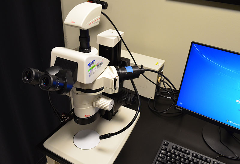
Leica Z16 Macroscope
- Objectives: 0.32xAchro,0.5xApo,1xApo, 2xApo
- Zoom range; 0.57x to 9.1x
- Magnification: 1xApo; 7.1x to 115x (2xApo total mag 230x)
- EL6000 Fluorescent Illumination
Filter Cubes
- A4 for UV, BP 470/40
- L5 for GFP, BP 527/30
- RFP, BP 605/75
- TX2 Texas Red, 645/75
Imaging Features
- Leica DMC 4500 Digital CCD camera, USB 3.0.
- 1280 x 960 pixel array
- Leica LAS Software
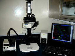
Olympus BX50
Observation Methods
- Brightfield
- Phase Contrast
- Differential Interference Contrast (DIC)
- Fluorescence
Digital Imaging
- Q-Imaging Retiga high resolution digital monochrome camera.
Computer and Software
- PC workstation with general office software and network connections
- Image Pro high-end image acquisition, processing, and analysis software
Filter Cubes
- Many filter cubes available upon request.
Objectives
- Many different objectives available upon request.
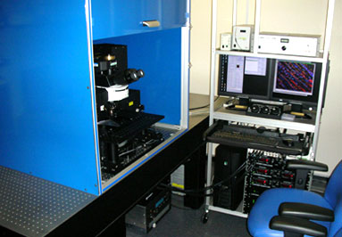
Prairie Ultima 2 Photon Microscope
- Transmitted and Fluorescence Illumination (Exfo Xcite)
- Upright Configuration
- Custom Heated Stage Insert
Laser Scanning Mode
- Galvo; for high quality imaging
- AOD; for fast imaging, e.g., 25 fps
- 4 Above Stage PMT Detectors (primary)
- 2 Substage Detectors (secondary)
Imaging Features
- Motorized stage: X, Y, and Z for optical sectioning
- 16 bit TIFF images
- 128, 256, 512, 1024, 2048 pixel resolution
- Frame Averaging
- Adjustable Dwell Time
- Advanced Time Series and Z-Sectioning
- Variable Zoom
- Atlas Mosaic Imaging
Coherent Laser
- Coherent Chameleon Ultra II
- Fast Tuning from 680nm - 1080nm
- Power output approximately 3000mW at 800nm
- Capable of exciting all fluorophores in the visible spectrum
Objectives:
- 10x/0.3 Dry WD 10mm,
- 20x/1.0 W Imm (XLUMPLN) WD 2mm,
- 40x/IR-2/0.8 W Imm WD 3.3mm,
- 60x/IR-2/0.8 W Imm WD 3.3mm
Spectra
- View PMT Detectors and Spectra
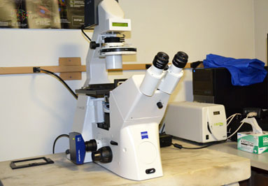
Zeiss AxioVert 200
Observation Methods
- Brightfield
- Phase Contrast
- Fluorescence (SOLA LED)
Digital Imaging
- Zeiss Mr3 camera (mono, 1388x1040 pixel array)
Computer / Software
- Dell Optiplex 790, Intel i5, 4 GB RAM, 2 19” Monitors
- Windows 7
- Zen Software
Objectives
- 5x/0.12NA A-Plan
- 10x/0.3NA EC Plan-Neofluar
- 20x/0.4NA LD Plan-Neofluar Corr
- 63x/1.4NA Plan-Apo Oil Imm
The Zeiss Inverted microscope is ideal for imaging assays prepared in well plates in either brightfield, phase contrast or fluorescence imaging modes. The fluorescent spectra of the filter cubes is optimized for DAPI, GFP, Cy3 and CY5 (blue, green, red and far-red).
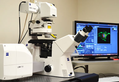
Zeiss LSM700
Widefield Visual Modes
- Bright Field
- DIC
- RGB Fluorescence: Exfo Xcite-120 Available Solid State LASERs
- 405nm
- 488nm
- 555nm
- 635nm
Detectors
- 2 Detectors with Spectral VSD (variable secondary dichroic)
- Transmitted Light Detector
Mounting Frames
- Universal Frame K for Slides
- Universal Frame KM for Plates
Imaging Features
- Observer Z1 Automated Stand
- Motorized: scanning stage for XY movements for tiling, Z for optical sectioning
- Single or multitrack image collection
- 8 or 12 or 16 bit pixel depth
- 128, 256, 1024, 2048 pixels per frame
- Zen 2012 Imaging Software for Acquisition and Analysis
Objectives
- Fluar 10x/0.5NA M27
- EC Plan-Neofluar 20x/0.5NA M27
- Plan-Apo 20x/0.8NA DICII
- Plan-Neofluar 40x/0.75NA DICII
- C-Apo 40x/1.2NA Water Imm DICIII
- iLCI Plan Neofluar 63x/1.3 Water Imm Corr M27