Light Microscopy/Digital Imaging
Light microscopy and digital imaging is integral to COHCCC’s research mission. The LMDI-SR provides a wide range of instruments that allow investigators to image numerous sample types in a variety of carriers such as slides, well plates, culture dishes, and live animals.
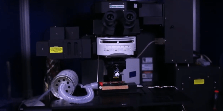
The LMDI-SR has 8 widefield microscope systems that have imaging capabilities in brightfield, fluorescence, differential interference contrast, phase contrast, and structured illumination (Apotome). The LMDI-SR has 5 systems that provide live-cell imaging services that include precise temperature control and CO2 delivery and a system that allows imaging under hypoxic conditions. The Zeiss confocal microscope systems (LSM700, LSM880, LSM900) are the most advanced available and are equipped with Airyscan super-resolution detectors that can increase the resolution and signal to noise ratio two-fold over confocal imaging. The LMDI-SR has two systems capable of 2-photon intra-vital imaging in live mice and rats. The Leica LMD7000 enables researchers to collect laser microdissected samples from slide mounted tissues, as well as live cells, which in turn can then be further analyzed via next generation sequencing, PCR, or mass spectrometry.
The LMDI-SR has added the Fluidigm Hyperion imaging mass cytometer to its services providing sophisticated multiparameter imaging data by ablating metal tagged antibodies from tissue samples. The LMDI-SR is directed by Dr Brian Armstrong who has extensive experience in light microscopic imaging and analysis and is supported by a highly trained staff (2 FTEs). The user base has access to the shared resource through iLab. In addition to extensive project-specific practical imaging training users also receive instruction in safety and the ethical collection of scientific imaging data. The LMDI-SR provides several professional software programs such as Image Pro Premier, AMIRA, Imaris, Autoquant, and Visiopharm that can be used to analyze and quantify imaging data, and the staff are well versed in the proper methods for handling large and complicated imaging data sets.
Note: This core is not supported by the National Cancer Institute-funded Cancer Center Support Grant.
Specific Aims of IMS-SR:
Aim 1. Provide support for COHCCC Members requiring expertise in light microscopy and digital image protocols.
Aim 2. Acquisition of imaging data for principal investigators.
Aim 3. Provide sophisticated image analysis and image presentation techniques to investigators.
Members Utilization by %Revenue 2017–21: 83.8 Total (25.7 MCBC, 10.1 DCT, 13.11 CI, 34.1 HM, 0.8 CCPS)
Publications by Members: 103, 39 with Impact Factor >10
Grants Supported: 143 Total (6 ACS, 8 CIRM, 8 DoD, 3 LLS, 64 NCI of 111 NIH (83 R01, 6 U01))
The objective of the Light Microscopy/Digital Imaging Core is to provide leading-edge microscopy systems and expertise to City of Hope investigators needing high-quality light microscope images and data. Microscopy and imaging are rapidly evolving fields that depend increasingly on expensive equipment and skills beyond the means of most individual laboratories. We seek to provide a range of research-grade light and confocal microscope platforms for simple observations and documentation suitable for publications or grant proposals. The core also makes available the latest research platforms for automated microscopy experiments, advanced observation techniques, and sophisticated digital image processing and analysis.
Services
- Instruction in general aspects of light and confocal microscopy
- Instruction in general aspects of digital imaging and image analysis
- Consultation on microscopy and imaging projects
Microscope Systems
- Widefield: __________7 Fluorescent Systems
- Live Cell Imaging: ____5 Zeiss Observers, 1 Nikon Biostation
- Confocal: __________ 1 LSM700, 2 LSM880 Airyscan
- 2-Photon: ___________1 Prairie Ultima with Coherent Chameleon
- LaserMicroDissection: _1 Leica LMD7000
- Digital Slide Scanner_Hamamatsu Nanozoomer HT2.0
- Fluidigm Hyperion IMC
Imaging Software for Image Quantification and Presentation
- Image Pro Premier
- Imaris 3D
- AMIRA 3D
- Autoquant Deconvolution
- Visiopharm
- Instruction in general aspects of light and confocal microscopy
- Instruction in general aspects of digital imaging and image analysis
- Instruction in operation of Light Microscopy/Digital Imaging Core equipment
- Consultation on microscopy and imaging projects


Michael Nelson M.S.
Research Associate II
mnelson@coh.org
626-256-4673, ext. 81259
Martha Salas
Research Associate I
masalas@coh.org
Contact the Team
The Hyperion imaging mass cytometer is part of Fluidigm's line of CyTOF systems which allows for tissue samples to be "imaged" and for the masses of metals to be quantified in relation to the tissue morphology at a resolution of one square micron.
The Leica laser microdissection microscope system has two widefield visual modes: bright field and RGB fluorescence/metal halide 120V.
The Leica MZ10F stereomicroscope features brightfield and fluorescence imaging, dual goosenecks for illumination and more.
The Leica Z16 macroscope offers objectives of 0.32xAchro, 0.5xApo, 1xApo and 2xApo, a zoom range from 0.57x to 9.1x and magnification of 1xApo and 7.1x to 115x (2xApo total mag 230x).
Olympus BX50 features observation methods: Brightfield, Phase Contrast, Differential Interference Contrast (DIC) and Fluorescence.
This microscope offers transmitted and fluorescence illumination (Exfo Xcite), upright configuration and a custom-heated stage insert.
Zeiss AxioVert 200 features observation methods like Brightfield, Phase Contrast and Fluorescence (SOLA LED).
This confocal microscope has various widefield visual modes, including Bright Field, DIC, RGB Fluorescence (Exfo Xcite-120 Available Solid State LASERs) and 405nm.
Zeiss LSM880 BRC offers various laser scanning modes, including Diode 405nm, Argon Laser 458nm, 488nm and 514nm, DPSS 561nm, and HeNe Laser 594nm.
Zeiss LSM880 Furth offers various laser scanning modes, including Diode 405nm, Argon Laser 458nm, 488nm and 514nm, DPSS 561nm, and HeNe Laser 594nm.
Zeiss Observer 7 BRC has various observation methods, including Brightfield, DIC and Fluorescence.
Zeiss Observer 7 features observation methods, such as Brightfield, DIC and Fluorescence.
Zeiss Observer II offers various observation methods, including Brightfield, DIC and Fluorescence.
Zeiss Observer Z1 offers observation methods, such as Brightfield Phase Contrast (5x only), DIC and Fluorescence.
In a given year, City of Hope conducts more than 400 clinical trials enrolling more than 6,000 patients.

City of Hope is focused on basic and clinical research in cancer, diabetes and other life-threatening diseases.


