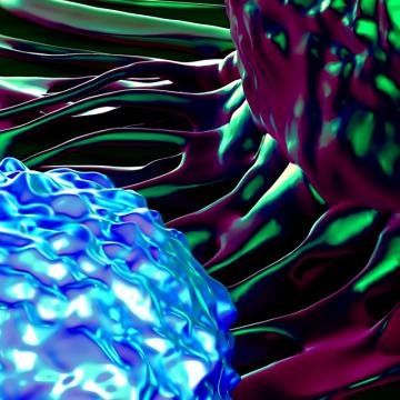
Electron Microscopy and Atomic Force Microscopy
City of Hope’s Electron Microscopy and Atomic Force Microscopy Core allows researchers to take sharper, closer looks at the building blocks of biological life. Electron microscopy (EM) and atomic force microscopy (AFM) provide imaging at a far greater resolution than light microscopy, allowing ultra-fine details of cells to be revealed and macromolecular structures to be solved. In addition, the EM/AFM Core is exceptionally well-equipped. The highly knowledgeable core personnel are pleased to help with experimental design, specimen preparation, data collection and image analysis.
The Electron Microscopy and Atomic Force Microscopy Core Facility is available to investigators wishing to define ultrastructural details in their experimental systems.
The core has three EMs and one AFM:
- Zeiss Sigma VP field emission scanning electron microscope equipped with multiple detectors (SE, BSE, STEM, and energy-dispersive X-ray spectroscopy for elemental analysis and mapping), with enhanced correlative microscopy capabilities and a Gatan 3View2XP serial blockface imaging device for serial blockface-scanning electron microscopy (SBF-SEM).
- FEI Tecnai 12 Twin transmission electron microscope equipped with a Gatan 894 Ultrascan 1000 CCD camera, Gatan cryo-stage/box, and Gatan tomography holder. In addition to traditional images, single-tilt tomography series and low-dose exposure images of specimens frozen in vitreous ice can be collected using the Tecnai 12.
- FEI Quanta 200 with environmental SEM capabilities.
- Bruker Multimode 8 atomic force microscope provides a means for imaging the surfaces of specimens at high resolution using physical probes. Cantilever holders allow for scanning in air and fluid. In addition, a ScanAsyst-HR high-speed Peak Force Tapping Scanner upgrade offers 20-times faster survey scans and six-times faster recording scans with no loss of resolution.
All instruments are user-friendly and can be operated by individual investigators following training.
In addition to providing conventional ultra-thin resin sections, the core facility provides sample preparation by high-pressure freezing and freeze substitution and for SBEM. Specimens can be analyzed by immunogold (pre- and post-embedding) labeling. A fully automated vitrification device, the FEI Vitrobot Mark IV, is available for plunge-freezing of aqueous (colloidal) suspension samples for particle analysis. Other types of sample preparation include negative staining, critical point drying, and shadow replicas. The PT-PC Ultramicrotome provides additional options for 3D reconstruction and correlative microscopy. The A3 specimen holder for SBEM extends analysis with the Gatan 3View2XP to individual cells and small cell populations, such as cell fractions from flow cytometry. Work stations and software are available for 3D visualization and tomographic reconstruction, segmentation, and particle analysis.
Preparation of specimens for electron microscopy
- Conventional chemical fixation and embedding of specimens in resin
- High-pressure freezing (HPF) and freeze substitution (FS) followed by resin embedding
- Ultra-thin sectioning of resin-embedded specimens
- Immunolabeling, with pre- and post-embedding methods
- Negative staining of small particles (e.g., viruses, nanoparticles and exosomes)
- Critical point drying and sputter coating for scanning electron microscopy
- Preparation of high-contrast specimens for serial blockface-SEM
- Plunge freezing for Cryo-EM (rapid freezing of samples in thin films on EM grids)
- Preparation of samples for AFM and EDS
- Correlative microscopy
Electron microscopy
- Collection of TEM, SEM (secondary and backscatter), Cryo-EM, STEM, and SBEM (serial blockface electron microscopy) images for 3D reconstruction, quantitative analysis, and segmentation
- Antigen localization by immune-labeling for EM (nanogold and other markers)
- Elemental analysis and elemental mapping
- Training in electron microscope operation and sample preparation
- Instruction in image analysis
- Correlative light and electron microscopy
Atomic force microscopy
- Collection of AFM data in a variety of modes
- Training in basic operation of the AFM





Contact the Team
Bruker Multimode 8 AFM provides a means for imaging the surfaces of specimens using a physical probe and nearly all the major scanning-probe-microscopy imaging methods, including contact and non-contact atomic force, lateral force, tapping (air), magnetic force, electric force microscopy, and ScanAsyst (Peak Force Tapping) techniques.
This coating system provides thermal evaporation of various metals. It is most often used for sputter coating SEM samples and depositing carbon onto TEM grids.
Freezing substitution is the follow-on procedure for high-pressure freezing. Specimens are fixed and resin-infiltrated at low temperatures providing higher quality structural preservation, including specimens suitable for immunolabeling for electron microscopy.
The HPF equipped with RTS allows ultra-rapid freezing of specimens. High-pressure freezing is superior to chemical fixation to preserve the native structure of cells and molecules.
This SEM is a versatile, high-performance (tungsten filament) scanning electron microscope with three operating modes (high-vacuum, low-vacuum and ESEM) equipped with a motorized stage and detectors for secondary and backscattered electrons.
This TEM provides several modes for directly imaging cell and macromolecular ultrastructure and can examine negatively stained specimens, cryo-specimens, and sections of resin-embedded specimens.
This fully automated vitrification device for plunge-freezing of aqueous (colloidal) suspensions provides a powerful means for determining the 3D structure of macromolecules and macromolecular complexes in their natural state.
The Gatan 3View2XP in-chamber microtome, combined with the Zeiss Sigma VP Field Emission Scanning Electron Microscope, produces 3D images in a fully automated, high-throughput and high-resolution fashion.
This instrument is available for identifying and mapping elements within specimens.
This instrument is available for specimen preparation allowing otherwise time-consuming sample preparation to be semi-automated and accomplished more quickly.
For rendering TEM carbon support films hydrophilic (negatively charged)
Critical-point drying provides better preservation of specimens for scanning electron microscopy.
This ultramicrotome (RMC Products, Boeckeler Instruments, Inc.) is ATUMtome equipped and used for producing serial sections for viewing in the Sigma VP Field Emission Scanning Electron Microscope and creating 3D image data.
For preparation of specimens for the scanning probe microscope
This versatile ultramicrotome is the workhorse instrument of the core. It provides sections of resin-embedded specimens prepared by conventional and cryo methods.
Commonly called a vibratome. The vibrating razor blade in the vibratome provides relatively thick sections (~100 microns) suitable for specialized electron microscopy techniques, including immuno-electron microscopy.
This FE-SEM is equipped with several detectors for secondary (SE) and backscattered (BSE) electrons.
In a given year, City of Hope conducts more than 400 clinical trials enrolling more than 6,000 patients.
Villamizar, O., Waters, S. A., Scott, T., Saayman, S., Grepo, N., Urak, R., Davis, A., Jaffe, A., & Morris, K. V.
Li, L., Tian, E., Chen, X., Chao, J., Klein, J., Qu, Q., Sun, G., Sun, G., Huang, Y., Warden, C. D., Ye, P., Feng, L., Li, X., Cui, Q., Sultan, A., Douvaras, P., Fossati, V., Sanjana, N. E., Riggs, A. D., & Shi, Y.
Patel, Z., Berlin, J., & Abidi, W.
Zhang, B., Nguyen, L., Li, L., Zhao, D., Kumar, B., Wu, H., Lin, A., Pellicano, F., Hopcroft, L., Su, Y. L., Copland, M., Holyoake, T. L., Kuo, C. J., Bhatia, R., Snyder, D. S., Ali, H., Stein, A. S., Brewer, C., Wang, H., McDonald, T., Swiderski, P. Troadec,E. Chen, CC, Dorrance, A. , Pullarkat, V., Yuan,Y.C., Perrotti, D., Carlesso, N., Forman, S. J., Kortylewski, M., Kuo, Y., Marcucci, G.
Shahin, S. A., Wang, R., Simargi, S. I., Contreras, A., Parra Echavarria, L., Qu, L., Wen, W., Dellinger, T., Unternaehrer, J., Tamanoi, F., Zink, J. I., & Glackin, C. A.
City of Hope is focused on basic and clinical research in cancer, diabetes and other life-threatening diseases.

