Chintan Parekh Lab Research
Defining the Earliest Stages of Human T Cell Development
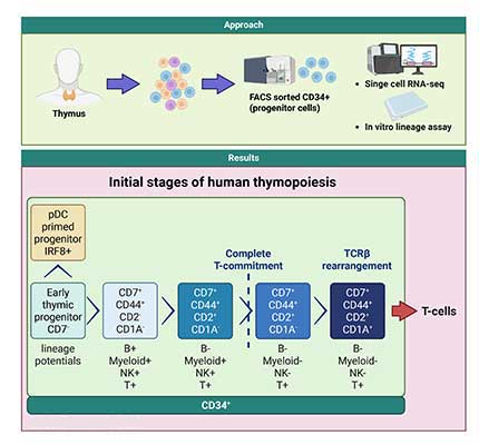
Understanding the stages of normal T cell development in humans is critical for elucidating mechanisms underlying T cell leukemogenesis, designing approaches to improve T cell reconstitution following bone marrow transplantation and enhancing the generation of T cell immunotherapies from stem cell sources. T cell development is initiated by the migration of multilineage blood cell progenitors (progenitors capable of giving rise to myeloid, T, B and NK lineage cells) from the bone marrow to the thymus. Within the thymus, these progenitors undergo a series of developmental stages where they lose other lineage potentials (T-lineage commitment) and ultimately give rise to mature T cells. However, the cellular and molecular events during the earliest stages of T cell development in the human thymus had been unclear.
Using single cell RNA-Seq (scRNA-Seq), we de novo reconstructed the T-cell developmental trajectory in the human thymus. We found that T cell progenitors are comprised of a continuum of previously undescribed developmental states. Bioinformatic analysis followed by functional studies revealed a species specific plasmacytoid dendritic (pDC) lineage primed IRF8 high progenitor population arising from the earliest T cell progenitors (ETP), findings indicative of an intrathymic dendritic specification pathway that diverges early from the canonical T-lineage differentiation pathway. CD2 expression defined stages of T-lineage commitment, with loss of B-potential preceding that of myeloid and NK potentials. ScRNA-Seq resolved the transcriptional landscape underlying the transitions between developmental stages and revealed key transcriptional differences in T cell progenitors between humans and mice.
We are now investigating scRNA-Seq inferred transcription factor networks via functional studies to elucidate the molecular mechanisms driving the earliest stages of human T cell development. A collaborative project between our lab and Gay Crooks’ lab at UCLA focuses on comparing these T cell developmental stages in the human thymus with those seen during T cell differentiation of pluripotent stem cells (PSC); the goal of this project is to enhance the generation of off the shelf T cell immunotherapies from PSC by identifying and optimizing the key biological processes needed for mimicking physiological T cell development during PSC differentiation.
The Role of BCL11B in Human T Cell Development
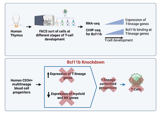
The initial stages of T cell development from blood cell progenitors involves two key processes, namely switching on T-lineage genes and switching off genes from other lineages (myeloid, B, NK lineages). The mechanisms driving these processes during human T cell development had been poorly defined. Through functional and next generation sequencing studies of human blood cell progenitors, we showed that the transcription factor BCL11B is needed for both inducing the expression of T-lineage genes as well as turning off genes from other lineages during human T cell development. ChIP-Seq profiles of progenitor cells from the human thymus demonstrated stage specific binding of BCL11B at multiple key T cell genes; expression of these binding targets was BCL11B dependent. We are now investigating the epigenetic mechanisms through which BCL11B mediates these two distinct processes with the goal of discovering T cell differentiation pathways that can be targeted to enhance T cell recovery post hematopoietic stem cell transplantation and increase the efficacy of T cell immunotherapies.
Improving the Efficacy of Immunotherapies
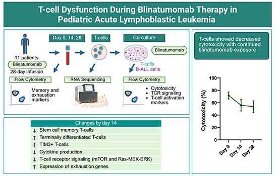
While the bispecific T cell engager Blinatumomab induces minimal residual disease (MRD) negativity in most adult and pediatric patients with B-cell acute lymphoblastic leukemia (B-ALL), loss or lack of response remains a clinical challenge. Blinatumomab is administered as a 28-day continuous infusion. Through high dimensional spectral flow cytometry, functional and RNA-Seq studies of T cells collected from pediatric patients with B-ALL at serial time points during Blinatumomab infusion, we found a decline in T cell cytotoxic function, TCR signaling, upregulation of the immune checkpoint molecule TIM3 and upregulation of an exhaustion gene signature by day 14 of Blinatumomab exposure. Our results support the study of treatment cycles with a shorter duration of continuous blinatumomab exposure. Gene expression analysis identified candidate T cell function genes for further investigation as potential targets for ameliorating Blinatumomab induced T cell exhaustion.