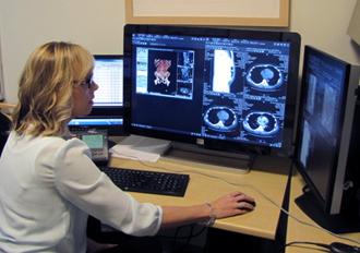
Clinical Care
3D Brain Imaging
Technology/services offered include:
- 3D Volumetric MRI
- Provides high resolution MR images of the brain
- Data can be imported into neuronavigation system
- Data can be used to perform volumetric analysis
- Beneficial in both intra-axial neoplasms in identifying macroscopic extent of lesion
- Added benefit in extra-axial neoplasms as it helps identify extension of skull base neoplasm
- CT and MR brain oncology assessment
- Brain tumor functional mapping
- Brain tumor mapping
- Brain tumor volumetric imaging
- Circle of Willis 3D volumetry
- Stroke evaluation and measurement
- 3D view of vascular and scalp
- ADC tumor assessment
- Tractography
- Fiber tracking can aid in identifying potential fibers that are either infiltrated by tumor or displaced, which can help identify potentially reversible and/or irreversible functions.
- This can help develop an optimal surgical resection plan
- Studies have shown increased frequency of negative resection margins when tractography is utilized.
3D Breast Imaging
3D tumor volumetric mapping allows surgeons and breast oncologists to follow the tumor pattern in dynamic MR imaging. It gives a better understanding of the tumor's aggressiveness and location and may be useful to help guide biopsies.
Technology/services offered include:
- MR breast tumor volume
- MR breast dynamics and functional mapping
- Fat measurement
3D Cardiac and Coronary Analysis
Technology/services offered include:
- Cardiac and coronary analysis
- 3D endovascular stent planning for aneurysm
- Evaluation of coronary stenosis
- Ejection fraction measurement
- Calcium scoring
- 3D cardiovascular anomaly assessment
- Assessment of flow dynamics and artificial valve
- Cardiac perfusion and bulls eye assessment
- 3D view of dilatation of aorta
3D Imaging Software
Imaging technologies used at the 3D Oncologic Imaging Center include:
- GE 3DAW Workstation
- 3D Vital images
- 3D Aegis
- iCAD
- DynaCAD
- 3D OsiriX
3D Liver Imaging
Hepatopancreatobiliary surgeries are among the most complicated procedures that need special presurgical measurement for optimal outcome. Quantitative presurgical segmental volumetric assessment allows surgeons to prepare for safety assessment whether the patient needs surgery, radiation or radio-embolization treatment.
3D advanced volumetric assessment gives a much stronger evaluation of the tumor compared to 2D estimation. Without 3D imaging, it is very hard for surgeons to understand the relationship between the tumor, the blood vessels, liver parenchyma or any other structure which is attached or close by the tumor. 3D volume quantification also allows surgeons to better communicate with patients about their surgical decision.
Technology/services offered include:
- 3D liver segmentation and voxel-based volume
- 3D portal circulation
- 3D MIP/IMIP imaging
- Liver transplant planning
- Liver tumor ablation 3D planning
- 3D view of liver vascular abnormality
- 3D presurgical planning for donors
- 3D liver tumor mapping with vascular details
3D Lung Imaging
Technology/services offered include:
- Lung cancer early detection screening
- Lung 3D volumetric segmentation and 2-D measurement
- 3D tumor volumetric assessment
- Lung airway flythru visualization
- 3D tracheo-bronchial abnormality
- Semi-automatic segmentation and characterization of nodule
3D Neck and Body Imaging
Technology/services offered include:
- 3D vascular abnormality in carotid and vertebral stenosis with plaque burden
- Stent planning
- PVD assessment
- Aneurysm assessment and measurement
- Carotid probing
- Lesion measurement
- Central line length and angle measurement
3D Pancreatic Imaging
- Presurgical planning for pancreatic tumor
- Pancreatic early detection imaging biomarker
- Pancreatic volumetric analysis on follow-up imaging
- Pancreatic 3D and pancreatic tumor vasculature
- 3D pancreatic histogram details
- 3D pancreatic textural analysis
3D Urology
Renal surgery planning is critical to decide whether patients need partial or radical nephrectomy (kidney removal). 3D advanced imaging is the best technology which provides high quality volumetric imaging with accurate quantitative measurement of tumor and kidney volume. It gives surgeons a better understanding of the size and location of a tumor and allows them to manipulate images while performing the surgeries.
Technology/services offered include:
- Renal tumor presurgical planning
- Merged multiphase CT imaging
- 3D tumor volumetric quantification
- 3D renal vascular evaluation
- 3D renal-tumor-vascular-ureter color mapping
3D Virtual Colonoscopy
3D virtual colonoscopy is a noninvasive procedure that provides a detailed image of the colon and rectum's interior lining. The procedure can detect and diagnose medical conditions, including precancerous polyps and cancerous tumors.
The procedure consists of two quick CT scans, one while lying on your back and one while lying on your stomach. Each scan takes less than a minute to complete.
Before the scan, your scan will be inflated with air or carbon dioxide to get a better image. You may experience a feeling or pressure or fullness, but there is typically no pain.
After the procedure, your radiologist will analyze the images and forward them to your physician, who will discuss the results with you.